As reported previously (1) and as shown in the figure below, ORP during 30-second epochs of sleep of any depth is not constant but is punctuated by events during which ORP rises to the wake level (e.g., > 2.0). Those lasting > 3 seconds are usually scored as arousals. More commonly, they are too brief or visually subtle to be scored as conventional arousals.
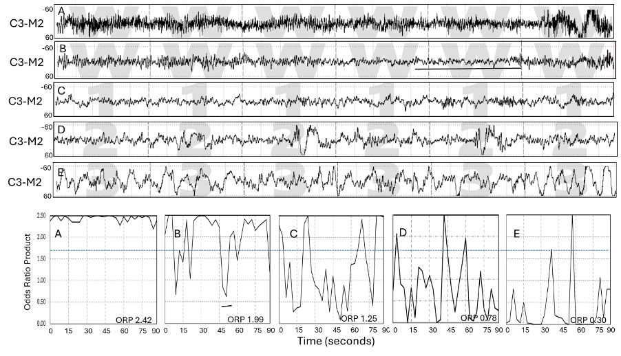
Figure: Illustrating Sleep intrusions during stage Wake (Panels A and B) and wake intrusions during NREM sleep of different depths: C, stage NREM-1, D, NREM-2, and E, NREM-3 in one normal sleeper. Each square at the bottom represents the time course of the odds ratio product (ORP) at 3-second intervals within 90 seconds (3 epochs), of which the middle epoch is the corresponding tracing above. Solid horizontal bars in the D and E EEG tracings represent the 3-second epochs with ORP>1.75. The dotted horizontal line at ORP 1.75 is the usual ORP cutoff between visually scored stages Wake and Sleep (2). Wake intrusions can be reported as the number of ORP spikes that exceed 1.75 or, more conservatively, exceed 2.0. From reference 1.
Wake intrusions are to be distinguished from evoked potentials in that they do not present as discrete waves of specific shapes. Rather, they reflect a change in the power spectrum of the EEG that is associated with a lighter sleep or awakening (increase in power in high frequencies and/or decrease in power in low frequencies (2,3). Wake intrusions likely represent arousal stimuli from somatic sources. Transient noise stimuli evoke similar ORP changes (4-6). When not associated with global arousals, they are very brief and strictly regional (i.e., there is little synchrony between intrusions in different brain regions), further supporting their origin from different somatic sources.
So far, information about wake intrusions has been derived only from central electrodes. The figure below from a study on 1368 patients with OSA (7) reflects what has been found in several studies. Their frequency (WII) varies greatly from 50 to more than 600 per hour (Figure). The frequency of wake intrusions directly impacts average ORP since the more frequently 3-second epochs with very high ORP occur in a 30-second epoch, the higher the average ORP. As a result, there is a strong correlation between WII and average ORPNR in the sleep study (Panel A, Figure, see also reference 8). On the other hand, the higher the average ORP is (lighter sleep), the more of the peripheral stimuli can reach the cortex and be detected. Since ORP-9 is a major determinant of average sleep depth, there is also a strong correlation between WII and ORP-9 (Panel B)..

WII is correlated with the conventional arousal/awakening index, but there is much scatter (panel C). The ratio of WII to the arousal index ranges from 5 to 20 in different people. WII also increases as a function of OSA severity, but there is again much scatter, with WII remaining low at very high AHI, and vice versa (Panel D). This may be relevant to the highly variable symptomatology in people with the same AHI. The weak association may be specific to central electrodes (i.e., OSA- related stimuli may project primarily elsewhere), or that much of the wake intrusions in these patients arise from other sources, or related to OSA but not to the events counted in the AHI (effort, hypoxemia, snoring, etc.).
The answers to these possibilities await further investigation. There was no association between WII and the PLM index in this study, which may, again, be specific to the central electrodes.
Panel F shows the results of multiple linear regression analysis of variables associated with WII in the same study (7). Average ORPNR was by far the dominant variable, followed by weak associations with ORP-9 and the arousal/awakening index.
The much higher frequency of WII than the arousal/awakening index suggests that WII may be a more sensitive indicator of sleep fragmentation. A recent study found that WII correlated with the frequency of insomnia symptoms in SHHS participants when the arousal/awakening index did not (Gratton).
Potential Role of Wake Intrusions in the Restorative Function of Sleep?
The discovery that perivascular spaces (PVSs) around cerebral blood vessels act as conduits (the glymphatic system) to circulate cerebrospinal fluid (CSF) within the brain parenchyma, to supply nutrients and remove waste products of brain activity (10), and the subsequent demonstration that sleep enhances the CSF flow through this system (11), provided the first concrete explanation of the mechanism of the restorative function of sleep.
The mechanism by which sleep promotes glymphatic drainage has been a subject of considerable debate. Several lines of evidence have recently converged on neurovascular coupling in the brain as the main driver of glymphatic drainage during sleep. Thus, glymphatic flow is markedly decreased during periods of increased neuronal activity (12-14), presumably because of an increase in brain cell size and compression of the glymphatic spaces. Intermittent brain activation is associated with fluctuations in CSF pressure, suggesting that intermittent brain activation results in corresponding compression and decompression of the perivascular spaces, promoting glymphatic flow (12).
This idea has received considerable support very recently in studies on mice, which documented sharp increases in norepinephrine levels at arousal associated with reciprocal changes in blood volume and consequent increase in CSF flow (15,16). This is a very relevant mechanism to sleep in humans since sleep is interrupted by arousals and awakenings at a rate that ranges from 12-36 per hour in healthy adults and up to 80/hour in patients with severe sleep fragmentation (17).
The neurovascular coupling mechanism is, however, a double-edged sword, since the exchange of some “dirty” CSF with “clean” CSF comes at the expense of more waste produced by the activated neurons. Given that one arousal or awake period can generate only one such exchange while the duration of the associated active state lasts several seconds (arousals) to minutes (awakening), the net effect on waste removal may be positive or negative depending on the duration of the active state and how much waste is removed during this single exchange. It may be expected, however, that briefer activations will be more effective than longer ones (1). Thus, the very brief wake intrusions may be the ideal form of brain activation for promoting waste clearance. Another advantage of wake intrusions is that they are regional. In this fashion, the pumping action and waste clearance of brain activation occur in all brain regions but not at the same time, thereby mitigating global arousals with their excitatory autonomic and cardiovascular consequences.
It is clear that wake intrusions offer a new, readily available, non-invasive approach to studying sleep fragmentation, as well as the potential role of an EEG biomarker in influencing waste clearance and, by extension, its possible role in cognitive and psychiatric disorders. The ability to obtain this information from a single EEG derivation makes it possible to evaluate the wake intrusion index in multiple regions from a single PSG with 4 to 6 derivations or in high density EEG.
Relevant References: